 EN
EN  DE
DE  ES
ES  PL
PL 


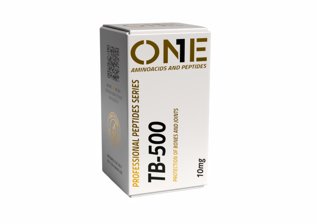
TB-500 5 mg – thymosin beta-4 analogue
TB-500 5 mg (chemical reagent, Thymosin beta-4 CAS No.: 77591-33-4) is a product intended for laboratory testing only – Research Use Only (RUO). The substance is not a medicinal product within the meaning of the Pharmaceutical Law Act of September 6, 2001, it is not a dietary supplement under the Food and Nutrition Safety Act, nor a veterinary medicinal product. The distribution is carried out in accordance with Art. 3a of the Pharmaceutical Law – exclusively for research purposes, including in vitro experiments on cell lines, studies on animal models and biophysical tests conducted by authorized laboratory personnel.
The history of the discovery of thymosin beta-4 (Tβ4) dates back to 1966, when Allan Goldstein and his team isolated a protein fraction with lymphocytopoietic activity from the calf thymus. The name “thymosin” referred to the source organ and suggested an immunological function. For the next quarter of a century, this peptide was classified as a factor regulating T cell maturation. It was only the work of Debora Safer and Maria Elzinga from New York (1991) that proved that Tβ4 is in fact the most abundant intracellular peptide sequestering monomeric actin – a discovery that completely reformulated the understanding of the biological role of this compound.
General description – chemical group and areas of scientific interest
In laboratory practice, this reagent is often compared with BPC-157 10 mg and PEG MGF 5 mgto assess differences in action profile across consistent study protocols.
TB-500 is classified as a synthetic peptide from the thymosin beta family – proteins with a mass of 4–5 kDa, which, despite their name, do not have a hormonal function and are not structurally related to thymosin alpha-1. The beta thymosin family includes three main isoforms in mammals: Tβ4 (dominant, present in virtually all types of nucleated cells), Tβ10 and Tβ15. All three isoforms bind G-actin, but differ in their affinity, tissue expression profile, and consequences of overexpression in culture models.
The scientific interest of TB-500 is focused on three research areas: (1) biophysics of actin polymerization and regulation of G/F-actin balance, (2) mechanisms of cell migration, including the dynamics of lamellipodial and filopodial structures, (3) modulation of repair processes in tissues in models of damage – with particular emphasis on models of cardiac ischemia-reperfusion in rodents and models of corneal damage.

What is TB-500? Biochemical definition and nomenclature problem
TB-500 is a synthetic peptide corresponding to the active region of thymosin beta-4, containing the sequence LKKTETQ (amino acid positions 17–23 in full-length Tβ4). Full-length Tβ4 is a polypeptide composed of 43 amino acid residues with a molecular weight of 4963.50 g/mol, acetylated at the N-terminus (Ac-Ser-Asp-Lys-Pro…).
The issue of nomenclature requires detailed explanation. In the literature on research reagents, the terms “TB-500” and “thymosin beta-4” are often used interchangeably, which leads to interpretation inaccuracies. From a strictly biochemical perspective, Tβ4 means a full-length 43-amino acid peptide, while the designation TB-500 refers to a synthetic fragment encompassing the active actin-binding region. This distinction has important methodological consequences: a peptide fragment may have a different binding affinity (Kd), a different proteolytic stability profile and a different ability to penetrate cell membranes compared to the full-length parent peptide. When designing experiments and interpreting results, it is important to precisely identify which peptide variant was used, and results obtained for Tβ4 should not be automatically extrapolated to TB-500 and vice versa.
The taxonomic affiliation of Tβ4 to the “thymosin” family is a historical artifact – it results solely from the isolation of this peptide from the thymus fraction (thymus) by Goldstein’s team. Functionally, Tβ4 is not a thymic hormone, but an actin-binding protein (ABP), whose intracellular concentration in proliferating cells reaches 300–500 µM, which makes it one of the most abundant cytosolic peptides.
Structure and physicochemical properties
Molecular architecture and conformation in solution
Tβ4 in its free form (unbound to actin) is intrinsically unstructured (intrinsically disordered protein, IDP). Circular dichroism (CD) and NMR spectroscopy studies showed the lack of a stable secondary structure in aqueous solution – the peptide adopts an extended random coil conformation with transient helical tendencies in the N- and C-terminal regions. Only in the presence of G-actin does Tβ4 undergo conformational ordering: the N-terminal helix (residues 4–16) binds the actin hydrophobic groove, the central LKKTETQ motif contacts subdomain 1, and the C-terminal helix (residues 30–40) covers subdomain 2 of the monomer.
This property – full structuring only in complex with a binding partner – places Tβ4 among the “folding upon binding” proteins, which has consequences for the kinetics of association: the lack of a rigid preformed binding structure allows for fast association (kon of the order of 10^7 M^-1 s^-1) at the expense of relatively low thermodynamic specificity.
LKKTETQ motif – amino acid residue analysis
The heptapeptide LKKTETQ (Leu17-Lys18-Lys19-Thr20-Glu21-Thr22-Gln23 in Tβ4 numbering) is the minimal sequence sufficient to bind G-actin. Leucine 17 interacts hydrophobically with the nonpolar pocket of subdomain 1. Two further lysines (Lys18, Lys19) form salt bridges with aspartate and glutamate residues on the actin surface. Threonine 20 and threonine 22, through the hydroxyl groups of the side chains, form hydrogen bonds that stabilize the orientation of the peptide in the binding groove. Glutamate 21, the only negatively charged residue within the motif, acts as a “conformational hinge” modulating the torsion angle of the main chain. Glutamine 23 closes the motif by participating in hydrogen cross-linking with the polar residues of actin subdomain 2.
Point mutations within LKKTETQ – described by Safer and Nachias’ team – showed that replacing Lys18 or Lys19 with alanine leads to a more than tenfold increase in the Kd of the complex with G-actin, confirming their essential role in the electrostatic interaction.
Physicochemical properties of the preparation
TB-500 in commercial form (One-Peptides, 5 mg) is supplied as a white to light cream-colored freeze-dried powder. The lyophilisate shows good solubility in deionized water, PBS (pH 7.4) and HEPES buffer (25 mM, pH 7.2) in the working concentration range of 0.1–100 µM. Storing the lyophilisate at -20 °C ensures stability for up to 24 months. After reconstitution, the solution must be stored at 2-8 °C; recommended consumption within 14 days. Division into aliquots immediately after dissolution reduces degradation resulting from repeated freeze-thaw cycles.
Mechanism of action at the molecular level
G-actin/F-actin balance – a central paradigm
Actin – one of the most conserved eukaryotic proteins – exists in the cell in two states: monomeric G-actin (globular, ~42 kDa) bound to ATP or ADP and polymeric F-actin (filamentous), forming double-stranded helical filaments. The G-actin → F-actin transition includes three phases: nucleation (formation of the nucleation trimer, the rate-limiting step), elongation (attachment of monomers to the (+) barbed end and (-) pointed end) and steady state (treadmilling – simultaneous polymerization at the (+) end and depolymerization at the (-) end).
Tβ4 and its synthetic fragment TB-500 act at the pre-nucleation stage: by binding G-actin in a 1:1 ratio, they lower the concentration of free monomers below the critical polymerization concentration (Cc, ~0.1 µM for the (+) end in the presence of ATP). However, they do not block the elongation of already initiated filaments – profilin, which also binds G-actin, catalyzes the exchange of ADP→ATP in the nucleotide pocket of the monomer and, via interactions with formins and VASP proteins, directs the monomer directly to the (+) end of the growing filament.
Tβ4/profilin competition – kinetic model
The ratio of Tβ4 to profilin concentrations determines the fate of the actin monomer. In the cytoplasm of unstimulated cells, Tβ4 quantitatively dominates over profilin (typical Tβ4:profilin ratio in thrombocytes is ~3:1), which maintains a large reserve of sequestered G-actin. Signaling stimulation (e.g. activation of the small GTPases Rac1 and Cdc42 by growth factors) recruits profilin to membrane nucleation complexes and locally tilts the balance towards polymerization. TB-500, added to an in vitro system, allows this ratio to be experimentally manipulated and the consequences on polymerization kinetics observed in assays with pyrene-labeled actin.
It is important that Tβ4 and profilin bind G-actin in different stereochemical configurations: Tβ4 blocks both the (+) and (-) ends of the monomer (total sequestration), while profilin binds the monomer only from the (-) end, leaving the (+) end capable of associating with the filament (partial sequestration, enabling directed polymerization). This fundamental mechanistic difference means that both proteins, although they compete for the same substrate, perform complementary functions in the cell.
Lamellipodium, filopodium and the Arp2/3 complex
The migrating cell forms two types of actin-rich projections on its leading edge: lamellipodium (wide, flat, composed of a branched network of filaments) and filopodia (narrow, finger-like, composed of parallel bundles of filaments). The organization of the lamellipodial network depends on the Arp2/3 complex – a seven-protein nucleator that binds the side of the existing filament and initiates the growth of a new branch at an angle of ~70°. Arp2/3 activation requires activating factors from the WASP/WAVE family, which in turn respond to signals from Rac1 (lamellipodia) or Cdc42 (filopodia, via mDia2 formins).
In this context, the role of Tβ4 / TB-500 is twofold: (1) maintaining the reserve of G-ATP-actin necessary for the rapid supply of nucleators – without an adequate pool of monomers, Arp2/3 is unable to support branch elongation, (2) modulation of the density of the filamentous network – increasing the concentration of Tβ4 shifts the balance towards depolymerization, reducing the density of the network, which paradoxically may increase the flexibility of the lamellipodium and facilitate cell navigation in the complex environment of the extracellular matrix. Microinjection experiments of Tβ4 into Swiss 3T3 fibroblasts (Carlier et al., 1996) showed changes in lamellipodium morphology dependent on the concentration of the injected peptide – low concentrations (<50 µM) increased protrusion dynamics, while high concentrations (>200 µM) led to lamellipodium collapsing due to excessive sequestration of monomers.
Applications in scientific research
Models of corneal damage – work by Sosne and colleagues
The cornea is an attractive tissue model for studying repair processes due to its optical transparency (enabling non-invasive imaging), lack of blood vessels, and a well-characterized population of epithelial cells with high migration potential. Gabriel Sosne’s team (Wayne State University) has published a series of papers examining the effects of Tβ4 in models of corneal epithelial damage.
In an alkaline corneal injury model in mice (NaOH 1 M, 30 s exposure), local administration of Tβ4 (0.1% w/v) to the corneal surface twice daily for 14 days led to accelerated reepithelialization compared to control (PBS), assessed by fluorescein staining and slit-lamp photographic documentation. Immunohistochemical analysis showed reduced expression of matrix metalloproteinases (MMP-2, MMP-9) in Tβ4-treated tissues and reduced concentration of pro-inflammatory cytokines (IL-1β, TNF-α) in corneal lysates, suggesting a two-pronged mechanism of action: pro-migratory (direct stimulation of epithelial migration) and modulating the inflammatory response.
In parallel in vitro experiments on human corneal epithelial (HCE-T) cells, Tβ4 increased migration in the scratch assay at a concentration of 1 µg/ml, and this effect was abolished by co-exposure with cytoclasin D (actin polymerization inhibitor, 0.5 µM), supporting a mechanistic link between Tβ4 promigratory activity and actin cytoskeleton dynamics.
Myocardial ischemia-reperfusion models – endpoints and quantitative parameters
Cardiac animal models are one of the most intensively researched areas of Tβ4 applications. In the work of Bock-Marquette and colleagues (Nature, 2004), administration of Tβ4 to C57BL/6 mice undergoing 30-minute ligation of the left descending coronary artery (LAD) followed by reperfusion led to a reduction of the infarct zone by ~40% compared to the control group (TTC staining, computer planimetry). Left ventricular ejection fraction (LVEF), assessed by echocardiography (40 MHz probe, M-mode) on the 28th day after the procedure, was 48 ± 3% in the Tβ4 group versus 35 ± 4% in the control group (p < 0.01).
The mechanism of the observed changes was attributed to the activation of ILK (integrin-linked kinase) by Tβ4, leading to phosphorylation of Akt (Ser473) and subsequent inhibition of caspase-9 in the apoptotic cascade. In additional experiments in isolated neonatal rat cardiomyocytes (NRCM), exposure to Tβ4 (100 ng/ml) reduced the percentage of TUNEL-positive cells after oxidative stress (H₂O₂, 200 µM) challenge from 38 ± 5% to 12 ± 3%.
In subsequent studies in a rat model (Wistar, LAD ligation 45 min), Smart and colleagues (Nature, 2007) showed that Tβ4 induces the mobilization of resident cardiac progenitor cells (c-kit+/Nkx2.5+) in the infarct border zone. This mechanism was associated with the pro-angiogenic activity of Tβ4 – increased neovascularization of the border zone confirmed by staining for CD31 (endothelial cell marker) and αSMA (pericyte marker), which indicated the formation of mature, stabilized vessels.
Scratch assay – methodological details
The scratch assay is the basic in vitro model for the quantitative assessment of cell migration. The experimental protocol includes the following steps: (1) culturing cells (e.g., NIH 3T3 fibroblasts, HUVEC endothelial cells, or HaCaT keratinocytes) to ~95% confluence in 12- or 24-well plates coated with type I collagen or gelatin; (2) serum starvation (medium with 0.5% FBS, 12 h) to synchronize the cell cycle and minimize the effect of proliferation; (3) performing a “scratch” using a sterile 200 µl pipette tip placed perpendicular to the bottom of the well – the movement must be uniform to ensure a reproducible gap width (~500 µm); (4) washing twice with PBS to remove detached cells; (5) adding medium with the tested peptide (TB-500 in the concentration range of 0.01–10 µM) and control; (6) photographic recording of the break at time points 0, 6, 12, 24 h using an inverted microscope (4x or 10x lens) with a CCD camera.
Quantitative analysis involves measuring the gap area at each time point using ImageJ software (Wound Healing Size Tool plug-in or manual segmentation). The result is expressed as a percentage of gap closure: [(field t=0 – field t=x) / field t=0] × 100%. Distinguishing the contribution of migration from proliferation requires a parallel experiment with preincubation with mitomycin C (10 µg/ml, 2 h before scratching) – cells treated with mitomycin retain their migratory ability but lose their division ability.
Biophysical tests – in vitro actin polymerization
TB-500 is used in biophysical tests of actin polymerization as a tool for controlled modification of the pool of available monomers. In a standard fluorescence assay, actin purified from rabbit skeletal muscles is labeled with pyrene (N-(1-pyrenyl)iodoacetoamide, Cys374 modification) – pyrenylactin fluorescence increases ~20-fold when switching from the G to the F form, which enables continuous monitoring of polymerization kinetics in real time. The addition of TB-500 to the reaction mixture at increasing concentrations (0.5–20 µM at a constant actin concentration of 5 µM) leads to a concentration-dependent delay of the lag phase (nucleation) and a decrease in the elongation rate, which allows Kd to be determined by fitting the Tb4 sequestration model.
Advanced variants of the test include the addition of profilin (at concentrations of 1–10 µM) to a mixture containing TB-500 and actin – such a three-component system allows for modeling the competition of both proteins for the monomer pool and determining the effective critical concentration of polymerization in conditions simulating cytosolic proportions.
Summary
TB-500, a synthetic peptide corresponding to the active region of thymosin beta-4 with the LKKTETQ motif, is a research tool with applications spanning actin cytoskeleton biophysics, cell migration biology, and experimental models of tissue damage. The mechanism of action is based on the sequestration of G-actin in a 1:1 ratio, modulating the G/F-actin balance in competition with profilin for the pool of monomers. In in vitro models (scratch assay, Matrigel tube formation test, pyrenylactin polymerization test) and in vivo models (cardiac ischemia-reperfusion models in mice and rats, models of corneal damage), changes in quantitative parameters dependent on the peptide concentration were observed.
All data quoted come from experiments on cell lines (NIH 3T3, HUVEC, RAW 264.7, HCE-T, NRCM) and animal models (C57BL/6 mice, Wistar rats) – extrapolation of the results to other organisms is not valid. The One-Peptides TB-500 5 mg product is a RUO class chemical reagent intended for research purposes only. Working with the preparation requires compliance with good laboratory practice (GLP) procedures and applicable ethical standards.
Parent category: Peptides for Regeneration organize RUO reagents associated with recovery-related research contexts.
FAQ
How is TB-500 different from full-length thymosin beta-4?
Thymosin beta-4 (Tβ4) is a full-length peptide composed of 43 amino acid residues (MW 4963.50 g/mol), while TB-500 is a synthetic fragment covering the active region of Tβ4 with the LKKTETQ motif. Both compounds bind G-actin, but may have different affinity (Kd), proteolytic stability profile and internalization capacity. In scientific publications, these terms should not be treated as synonyms – precise identification of the peptide variant used is a condition for the correct interpretation of the results.
What cell lines are most often used in studies with TB-500?
Previous publications most frequently used: NIH 3T3 and Swiss 3T3 fibroblasts (migration tests, microinjections), HUVEC endothelial cells (Matrigel angiogenesis tests, scratch assay), RAW 264.7 mouse macrophages (NF-κB modulation), HCE-T human corneal epithelial cells (corneal damage models) and NRCM rat neonatal cardiomyocytes (stress models). oxidative).
What controls should be included in the scratch assay with TB-500?
A properly designed experiment requires: negative control (medium with carrier without peptide), proliferation control (preincubation with mitomycin C, 10 µg/ml, 2 h), a series of peptide dilutions (minimum 3–4 concentrations, e.g. 0.01; 0.1; 1; 10 µM) and a minimum of three biological repeats. Photographic documentation should include at least 3 fields of view per well at each time point.
Under what conditions should TB-500 be stored after reconstitution?
The solution after reconstitution in deionized water or PBS (pH 7.4) should be stored at 2-8 °C, divided into aliquots to avoid multiple freeze-thaw cycles. The recommended storage period after dissolution is up to 14 days. The unopened lyophilisate is stable for up to 24 months at -20 °C.
How to distinguish the migratory effect from the proliferative effect in experiments with TB-500?
The distinction requires a parallel experiment with a proliferation inhibitor. Mitomycin C (10 µg/ml, preincubation 2 h) alkylates DNA and blocks cell division, without affecting the migratory ability. Comparison of the rate of gap closure in the scratch assay between cells treated and untreated with mitomycin C in the presence of TB-500 allows the separation of both components. Alternatively, BrdU or EdU labeling allows quantification of proliferation in the gap closure zone.
Scientific sources
- Safer D, Elzinga M, Nachmias VT (1991). Thymosin beta 4 and Fx, an actin-sequestering peptide, are indistinguishable. Journal of Biological Chemistry. (PMID: 1999398; actin-binding peptide characterization)
- Bock-Marquette I et al. (2004). Thymosin beta4 activates integrin-linked kinase and promotes cardiac cell migration, survival and cardiac repair. Nature. (PMID: 15565145; effect of Tbeta4 on heart cell migration and survival)
- Sosne G et al. (2010). Biological activities of thymosin beta4 defined by active sites in short peptide sequences. FASEB Journal. (PMID: 20179146; thymosin beta-4 active regions)







Reviews
There are no reviews yet.